
Getting It Wrong In Clay Before Getting It Right In Neck Dissection
Ka Wai Chung (Jacky) - BSMS JTG Rep
For most medical curriculum, anatomy is taught in 2 modes, on textbooks where we trace what someone else has drawn, or in dissection where we observe structures that have are displayed. Neither ask for a decision.
Watching my first neck dissection, I realised the gap between knowing anatomy and understanding it. I could identify the broad landmarks – sternocleidomastoid, position of the common carotid artery and internal jugular vein, and roughly where levels I to V sat in relation to them. But when the surgeon began working around the structures along fascial planes to sperate nodal level from the next, I was lost. The names existed somewhere in my memory but how they sat relative to each other in a live surgical field with anatomical variations was something I had never truly built a picture of.
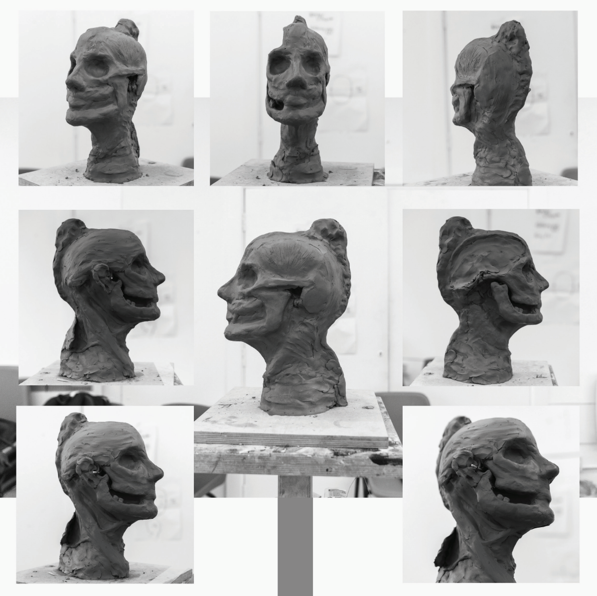
Most of us have sat with a pencil and worked through the neck triangles, the lymph node levels, the fascial layers. It consolidates ideal anatomical locations, but it remains two-dimensional. Sculpting is different. When you build a neck in clay, you are forced to make anatomical decisions in three dimensions. You can’t define level IIa without committing to the boundaries created by the internal jugular vein, the spinal accessory nerve, and the posterior belly of the digastric. You can’t appreciate why the marginal mandibular branch of the facial nerve is at risk in level I dissection until you’ve placed the submandibular gland and traced what runs above it. And crucially — unlike a cadaver or a diagram — you can keep going. You can refine a border, bury one structure beneath another, and interrogate the relationships between them until they make sense. The anatomy becomes something you explore rather than observe. You begin to think about operative zones and surgical planes, not just names on a page.
As a medical student with little formal dental education, the exercise brought an unexpected dividend too. Building the mandible in clay — establishing the condylar head and its relationship to the glenoid fossa, the sweep of the ramus down to the body, the alveolar process housing the dentition — gave me a structural picture I had never properly formed. For the first time I could appreciate how the occlusal plane and the alignment of the dental arches are direct consequences of the underlying bony architecture, and why a mandibular resection doesn’t just change a patient’s appearance but fundamentally disrupts their bite and function. It gave me a way into conversations about occlusion and reconstruction that I wouldn’t otherwise have had standing on the medical side of an OMFS ward round.
Sculpting alone didn’t close the gap entirely. Attending FlexFacs, the online teaching series run by BAOMS where consultants walk through facial planes using diagrams and clinical images, added another layer. Seeing those planes illustrated and discussed by surgeons who operate in them daily helped translate the three-dimensional picture I was building into something clinically grounded. The two experiences complemented each other — sculpting gave me the architecture, FlexFacs showed me how surgeons think about navigating it.
Together they shifted my understanding in a way that textbooks and dissection room prosections alone hadn’t managed.
For medical and dental students considering OMFS, getting ahead of the anatomy early matters. The specialty operates in one of the most complex anatomical regions in the body, and the gap between textbook knowledge and operative understanding is wide. Cadaveric dissection is irreplaceable when it’s available. When it isn’t, you have to be creative.
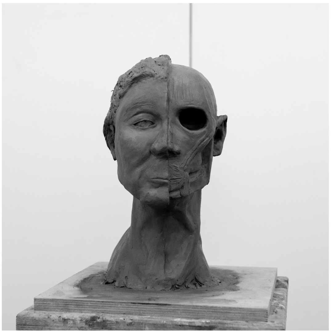
Last August I spent five days on a portrait sculpture course at Phoenix Brighton with Hazel Reeves MRSS. By the end I had a life-size clay bust, half surface anatomy, half dissected — and a clearer picture of the craniofacial region than I had managed to build in two years of medical school.
You don’t need a formal course. You need clay, some reference material, and the willingness to get it wrong and start again. Which, come to think of it, is reasonable preparation for surgical training.
Click here to see Jacky’s portfolio
