

JTG 2025 Poster Gallery
Orthognathic Surgery Outcomes: A Regional Unit’s BAOMS Audit Findings
Introduction
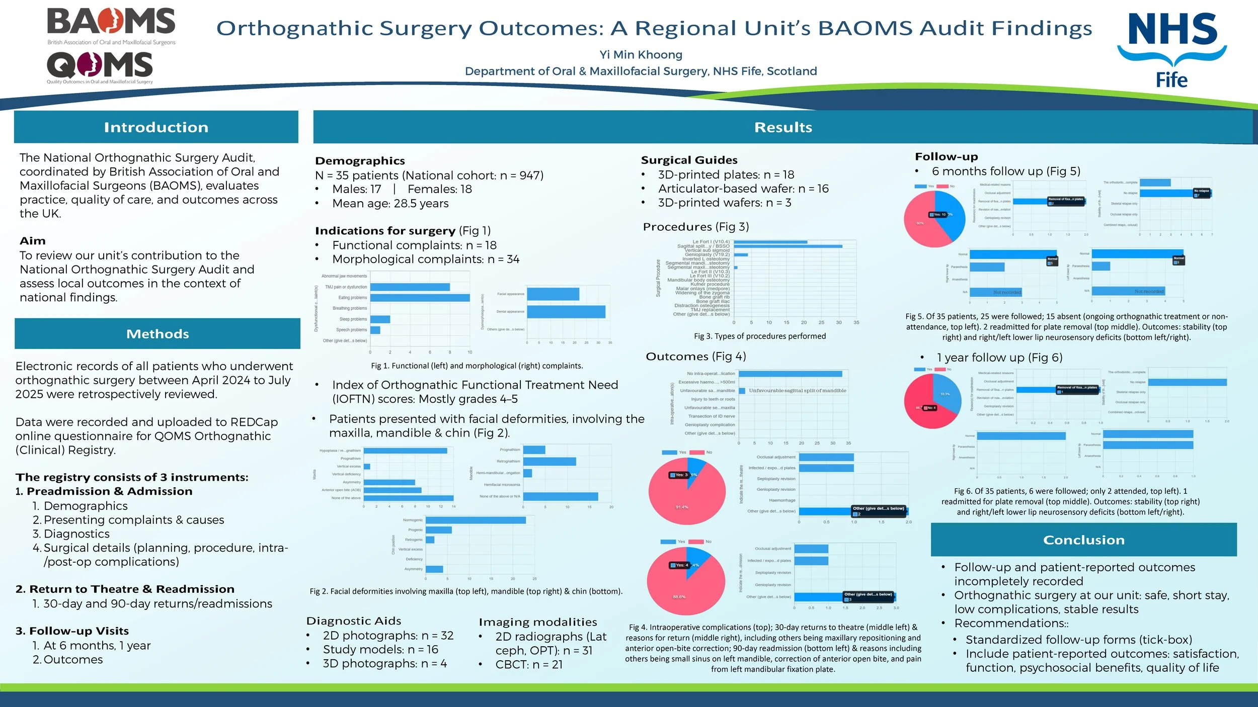
The National Orthognathic Surgery Audit conducted by British Association of Oral and Maxillofacial Surgeons (BAOMS) aims to assess practice, quality of care, and outcomes throughout the UK. This retrospective audit presents findings from a regional maxillofacial unit, which contributed 35 patients to the national cohort of 947 from April 2024 to July 2025.
Methods
A review was conducted of all patients who underwent orthognathic surgery at our unit during this period. Data was collected on demographics, presenting complaints, diagnoses, treatment plans, surgical procedures, and postoperative outcomes.
Result
The cohort included 17 males and 18 females, with a mean age of 28.53 years. Indications encompassed functional issues (e.g., masticatory difficulties, TMJ dysfunction), and morphological deformities (facial, dental appearance). IOFTN scores were predominantly 4–5, reflecting high need.
The majority underwent combined procedures, most commonly Le Fort I osteotomy and bilateral sagittal split osteotomy, with a smaller proportion receiving genioplasty. Planning methods included both traditional and digital approaches, ranging from standard articulator-based techniques and wafers to advanced 3D planning with 3D-printed wafers.
The median inpatient stay was 1 day (range 0–2), with 10 patients discharged within 24 hours. Intraoperative complications were uncommon, with 2 cases of unfavourable sagittal split of mandible. A minority (5 patients) returned to theatre within 90 days, primarily for fixation plate-related problems and occlusal adjustments.
At follow-up, outcomes were stable with minimal relapse rates and acceptable neurosensory deficit. One readmission occurred within 1 year for plate removal. Documentation of follow-up and patient-reported outcomes was inconsistent across cases.
Conclusion
Orthognathic surgery at our unit demonstrates safe practice, short inpatient stay, low complication rates, and stable results. Follow-up documentation could be improved using a standardized tick-box, and patient-reported outcomes (e.g., satisfaction, functional and psychosocial benefits, quality of life) should be incorporated to better capture treatment impact.

Literature review: Intraosseous Hemangiomas of the facial bones: Diagnosis and management
Introduction
Intraosseous hemangiomas of the facial bones are rare benign tumours which are vascular in nature. This vascularity makes management difficult and often require a multidisciplinary approach for management. I have reviewed published literature to outline their presentation, the diagnostic tools available and management options.
Method
Articles were searched for by using pubmed medical database and reviewing articles references for similar articles.
Results
Intraosseous haemangiomas can present as painless bony lumps on the face or as space occupying lesions which compress structures, causing associated symptoms. These can include proptosis, facial deformity and dysethesia. Rarely they are painful. They can have differing vascular pressure, with a small number presenting as a pulsatile mass with bruit. There is no clear consensus on why they form, with a history of trauma and congenital reasons being suggested.
They are often first noted on CT imaging, showing heterogeneous honeycomb appearance. The benefit of MRI in diagnosis is disputed, as they can present as either high, low or mixed intensity on T1. However, MRI is useful to review vascularity within the lesion. Incisional biopsies have a limited role due to bleeding risk.
If no functional or aesthetic concern they can be managed conservatively. Non-surgical options attempted include needle aspiration, sclerotherapy and radiotherapy, all showing mixed results.
Surgical options are complicated by bleeding risks. This can be managed by removal ‘en-bloc’ to eliminate the need to enter the lesion, whilst pre-surgery embolization has also be utilised. Partial lesion removal has also been attempted, to restore function only.
Conclusion
Intraosseous hemangiomas of the facial bones are rare and limited published evidence exists, mainly case reports. There is not yet a consensus on the most useful diagnostic tools or their management. Each lesion presents its own challenges in removal and reconstruction dependent on size and location.